Breast MRI for Early Detection of Breast Cancer
Breast magnetic resonance imaging (MRI) is a procedure being studied more frequently for its role in detecting breast cancer, including the diagnosis of ductal carcinoma in situ (DCIS) and contralateral breast cancer (breast cancer in the opposite breast) at the time of a first breast cancer occurrence. DCIS is a pre-invasive form of breast cancer. Nearly all invasive breast cancers are thought to develop from precancerous lesions (areas of abnormal tissue) seen in DCIS. The high-grade form of DCIS grows quickly and is more likely to develop into invasive breast cancer.
Mammography is the standard method for diagnosing DCIS, which accounts for 20% of diagnosed breast cancers. Although the early results of breast MRI studies are encouraging, breast MRI should not be substituted for mammography for women at average risk for breast cancer. However, it may be an additional tool to screen for breast cancer in women at high risk for developing the disease.
Recommendations for breast cancer screening
New guidelines from the American Cancer Society recommend breast MRI and mammography yearly beginning at age 30 for women at high risk for breast cancer. This population includes women who meet at least one of these criteria:
- known BRCA1 or BRCA2 gene mutation
- strong family history of breast or ovarian cancer
- a 20% or greater lifetime risk of breast cancer (this can be calculated using a scientific tool that calculates a person's lifetime risk of developing breast cancer)women who have received radiation therapy to the chest between the ages of 10 and 30
- women with a first-degree relative with the BRCA1 or BRCA2 gene mutation who have not had testing themselves
- women who have, or may have, a family history of a cancer syndrome that increases their risk of breast cancer
Comparing breast MRI and mammography
- MRI and mammography both take pictures of the breast, but in different ways. Here are some differences between breast MRI and mammography.
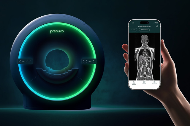
- An MRI uses magnets that emit radio waves to produce a three-dimensional view of the breast and the underlying structures and vessels. Mammography uses low doses of radiation (x-rays) to produce a one-dimensional image of the breast.
- Breast MRI is a more sensitive test than mammography, especially when given with a contrast medium (a special dye). The dye makes the cancerous area of the breast appear much brighter, but it may cause many areas of the breast that do not have cancer to appear abnormal, causing an increased number of false-positive test results. This may cause unnecessary biopsies and increased anxiety for many women.
- Breast MRI cannot visualize calcifications (calcium deposits) and microcalcifications (irregular deposits of calcium) that typically surround DCIS lesions. Mammography, on the other hand, can visualize calcium deposits accurately.
- A positive finding on an MRI requires a follow-up biopsy to confirm whether a suspicious finding is cancerous. Many facilities are not equipped to perform biopsies with MRI guidance. Women may have to repeat MRI testing with a follow-up MRI-guided biopsy at another facility that is equipped to perform the procedure. A positive finding on a mammogram also requires a follow-up test, such as diagnostic mammography, ultrasound, and/or biopsy, but these tests do not require the specialized technology of an MRI-guided biopsy.
- Breast MRI may be better able to visualize dense breast tissue compared with mammography, and may be better for younger women with a high risk of breast cancer.
- A breast MRI is expensive, costing anywhere from $800 to $2,000, compared with a mammogram, which costs anywhere from $85 to $150. Most insurance plans and Medicare cover a mammogram every 12 months, and women may be responsible for a small copayment. Not all insurance companies provide reimbursement for breast MRI for women at average risk for breast cancer (breast MRI may be covered for women at high risk for breast cancer). Medicare allows for "reasonable and necessary" breast MRI if performed on equipment approved by the U.S. Food and Drug Administration.
- Currently, no formal practice standards exist for breast MRI as they do for mammography. Training and accreditation (a process to determine if applicable standards are met) of MRI facilities is needed to ensure practice standards remain consistent from one facility to another. The American College of Radiology is in the process of developing such standards.
Questions to Ask Your Doctor
Breast MRI has certain limitations and is not intended for all women. Discuss any concerns about early breast cancer detection with your doctor, and consider asking the following questions:
- What is my risk of developing breast cancer?
- Based on my risk of developing breast cancer, do you recommend any additional screening tests, such as breast MRI, besides annual mammography?
- If I have never had a mammogram, should I start my screenings with an MRI?
- What are the risks and benefits of having a mammogram?
- What are the risks and benefits of having a breast MRI?
- Before I schedule my MRI, should I contact my insurance company to see if it is covered?
- What is the next step if something is suspicious on the mammogram?
- What is the next step if something is suspicious on the MRI?
This information was reprinted with permission. © 2008 American Society of Clinical Oncology. All rights reserved. www.Cancer.net


